The Transylvanian area of Romania boasts a rich fossil record of dinosaurs, which lived on an island (Haţeg Island) during the very end of the Cretaceous. Many of these are dwarfed in body size or exhibit other unusual features thought to be linked to their insular habitat. One of the most common of these dinosaurs is the rhabdodontid ornithopod Zalmoxes, an herbivorous taxon that has been found at many Upper Cretaceous sites across Transylvania. Our collaborative fieldwork has uncovered several new Zalmoxes specimens from the Nălaţ-Vad (= Vadu) locality, a site along the Râul Mare River in the Haţeg Basin that dates to the “middle” to late Maastrichtian. These include a partial associated skeleton, along with various isolated bones from several additional individuals. We catalog and describe these specimens here, and compare them to other Zalmoxes fossils from Romania. They provide further evidence that Zalmoxes was one of the most common vertebrates in the latest Cretaceous of Transylvania, and add to the unusual fossil record of Nălaţ-Vad, which has yielded a much greater number of associated skeletons than other Transylvanian localities. Some of the Nălaţ-Vad specimens possess features characteristic of the type species, Z. robustus, whereas others exhibit features diagnostic of the larger and stockier Z. shqiperorum, indicating that these species were locally sympatric, as has been demonstrated at other Transylvanian sites. The specimens span much of the size range known from Zalmoxes, as well as the spatial and temporal extent of the Nălaţ-Vad locality, suggesting that this taxon flourished until near the end of the Cretaceous.
INTRODUCTION
The Transylvanian region of Romania is one of the few places in the world that yields fossils of dinosaurs and other vertebrates from the final few million years before the end-Cretaceous mass extinction (Weishampel et al., 2004; Brusatte et al., 2015; Csiki-Sava et al., 2016). These Romanian dinosaurs were highly unusual compared with species from other parts of the globe, as many were dwarf forms or had evolved bizarre morphological features, most likely because of their insular habitats (Nopcsa, 1914; Weishampel et al., 1991; Benton et al., 2010; Weishampel and Jianu, 2011; Brusatte et al., 2013a; Csiki-Sava et al., 2015). The great majority of the Romanian dinosaurs come from the Haţeg Basin in western Transylvania, a fossil-rich area that was originally explored by the pioneering paleobiologist Ferenc Nopcsa in the early 20th century (e.g., Nopcsa, 1915, 1923), and then later by international teams led by Dan Grigorescu (Bucharest) and David Weishampel (United States) in the 1980s and 1990s (e.g., Weishampel et al., 1991).
More recently, our team has been studying the vertebrate paleontology and geology of the Transylvanian continental uppermost Cretaceous, building on the earlier work of our Romanian team members Mátyás Vremir and Zoltán Csiki-Sava. Our focus has been on collecting new specimens from the Haţeg Basin and exploring other nearby sedimentary basins, in order to better understand the latest Cretaceous Romanian faunas and what they reveal about how dinosaurs adapted to insular environments and were affected by the end-Cretaceous extinction. Our team includes scientists from the Transylvanian Museum Society (Cluj-Napoca, Romania), the University of Bucharest (Romania), the “Ioan Raica” Municipal Museum (Sebeş, Romania), the American Museum of Natural History (New York), and the University of Edinburgh, often in collaboration with many other researchers from various institutions. We have been working together since 2010 and undertaking annual field expeditions during that time, and the Romanian members of the team have also been conducting regular fieldwork throughout the years.
Thus far, some of the more important results of our collaboration have been the initial report (Csiki et al., 2010a) and monographic description (Brusatte et al., 2013a) of Balaur bondoc, an aberrant double-sickle-clawed dromaeosaurid that was discovered by Mátyás Vremir in 2009; studies on the origin of the unusual Romanian dwarfed dinosaur faunas (Vremir et al., 2014) and their relevance for understanding the end-Cretaceous extinction (Csiki-Sava et al., 2016); and descriptions of new infant dinosaur (Brusatte et al., 2013b) and midsized pterosaur (Vremir et al., 2015) material.
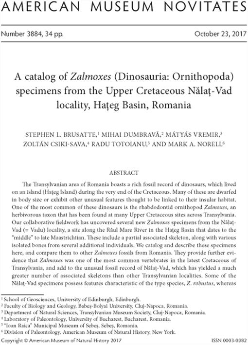
As part of our field project we have been making frequent trips to the Nălaţ-Vad (= Vadu) locality, a site along the Râul Mare River in the Haţeg Basin (fig. 1), where fossiliferous exposures of “middle” to upper Maastrichtian rocks extend for approximately 500 meters of riverbed. Fossils from along the Râul Mare valley (between Orlea, Vadu, Toteşti and Unciuc) were first noted by Nopcsa (1905), but more significant vertebrate fossils from Nălaţ-Vad were reported only a century later by Smith et al. (2002). Over the 15 years since, a large number of vertebrate (including dinosaur) bones, teeth, eggs, and nests have been discovered at this locality (e.g., Van Itterbeeck et al., 2004; Godefroit et al., 2009; Csiki et al., 2010b; Grigorescu et al., 2010; Wang et al., 2011; Csiki-Sava et al., 2016).
FIGURE 1.
A, Location of the Haţeg area in Romania and B, simplified geological map of the Haţeg Basin (modified from Csiki, 2005; Grigorescu and Csiki, 2008). Locality names in blue mark latest Cretaceous (Maastrichtian) fossiliferous localities, with Nălaţ-Vad locality highlighted in capital letters. Abbreviations: D-C F, outcropping area of Densuş-Ciula Formation; RMb, outcropping area of the informal “Râul Mare beds”; SF, outcropping area of Sînpetru Formation.

We have discovered several new specimens of the rhabdodontid ornithopod dinosaur Zalmoxes from Nălaţ-Vad. Zalmoxes is one of the most common vertebrates in the latest Cretaceous of Romania and is often considered to be an island dwarf (e.g., Weishampel et al., 2003; Benton et al., 2010), although this is debatable (see review in Ősi et al., 2012; Prondvai, 2014). Two sympatric species of Zalmoxes were identified by Weishampel et al. (2003) in their original description of the genus. However, some portions of the Zalmoxes skeleton remain unknown, and there are still open questions about its taxonomy and systematics, as well as its body size. In this short report we describe and figure the Zalmoxes specimens that we have recovered from Nălaţ-Vad, to provide a catalog of the fossils of this important dinosaur that have stemmed from our field project at this particular locality.
MATERIALS AND METHODS
All specimens illustrated here were mechanically prepared using a CP (Chicago Pneumatic) 9361 air scribe equipped with either a chisel or needle point and a Ken Mannion prototype ST model air scribe, operated at a pressure of 116 PSI. Some specimens were additionally prepared chemically via immersion (1 hr) in a 3% weight-by-volume solution of acetic acid (CH3COOH) in water. After preparation, the specimens were coated with a 5% weight-byvolume solution of Paraloid B72 dissolved in acetone in order to stabilize and preserve the bone surface. 3D models of the right scapula LPB (FGGUB) R.2164 (see fig. 12) and left femur LPB (FGGUB) R.2408 (see fig. 16) were constructed using digital photogrammetry, following the methodology described by Mallison and Wings (2014). A total of 70 photographs in two orientations were acquired by placing the specimens on a manual turntable in 5° increments. The textured surface models were reconstructed using Agisoft Photoscan PRO. After reconstruction, the models were saved as 3D interactive PDFs using Adobe Acrobat X Pro.
SYSTEMATIC PALEONTOLOGY
Dinosauria Owen, 1842
Ornithischia Seeley, 1887
Ornithopoda Marsh, 1881
Iguanodontia Sereno, 1986
Rhabdodontidae Weishampel et al., 2003
Zalmoxes Weishampel et al., 2003
Nomenclatural Note: In their original description of Zalmoxes, Weishampel et al. (2003) considered the genus to contain two species that are both present in the uppermost Cretaceous of Romania: the type species Z. robustus (originally described by Nopcsa [1902] as Mochlodon robustus) and the newly erected Z. shqiperorum. While Z. robustus was considered to be represented by abundant specimens from across Transylvania, Z. shqiperorum was originally based on a single incomplete adult specimen (the holotype) and a more limited set of referred fossils (Weishampel et al., 2003).
More recently, Godefroit et al. (2009) described a more complete skeleton of Zalmoxes shqiperorum from the Nălaţ-Vad locality (UBB NVZ1; see fig. 22) that includes dissociated elements of the skull together with elements of the axial and appendicular skeleton (table 1). Additionally, Godefroit et al. (2009) also referred to Z. shqiperorum a second, less complete partial postcranial skeleton (UBB NVZ2: scapula, humeri, femora) from Nălaţ-Vad, along with further isolated rhabdodontid remains from Nălaţ-Vad (dorsal and caudal vertebrae, humerus, ulna, femora, tibiae) and the Sibişel Valley (UBB SPZ-2: associated sacrum and pelvic girdle from La Scoabă locality; see Csiki-Sava et al., 2016). However, no positive evidence was presented to support these specific referrals. It is thus unclear whether all this rhabdodontid material can be referred to Zalmoxes shqiperorum, especially in the case of isolated skeletal elements that overlap neither with the holotype nor with the more complete referred specimen UBB NVZ1 (e.g., humeri, tibiae). Furthermore, some of the elements assigned by Godefroit et al. (2009) to Z. shqiperorum (e.g., UBB NVZ9, an isolated right femur; UBB NVZ1-40, a basioccipital [not to be confused with the left partial quadrate bearing the same number in Godefroit et al., 2009: fig.5]; possibly also UBB NVZ3, an isolated left tibia) bear features that cannot be reconciled with the genus Zalmoxes, and might instead belong to the hadrosauroid Telmatosaurus.
Based on this new material (the referral of some of which is contentious), Godefroit et al. (2009) provided a revised diagnosis of the genus Zalmoxes and the species Z. shqiperorum, and commented on the diagnosis of Z. robustus. Unfortunately, these diagnoses are hampered by some unavoidable difficulties. First, except for a few cases of positive and well-documented field association (e.g., Nopcsa, 1904, 1928; Weishampel et al., 2003; Godefroit et al., 2009; Csiki et al., 2010c; Botfalvai et al., 2017), the vast majority of the previously known Zalmoxes fossils have been disarticulated and/or isolated, making their referral to one or the other of the two species largely conjectural. There are many features that may be diagnostic of either Zalmoxes or one of the two species, but these cannot be assessed in close relatives (such as Rhabdodon) and/or in one of the two Zalmoxes species because of a lack of comparable bones. It is also unclear how some of these features may be related to individual variation or ontogeny, although there is limited evidence that specimens of similar ontogenetic stage are known for both species, indicating that one is not the juvenile of the other (Ősi et al., 2012; Prondvai, 2014). Alternatively, it is also conceivable that some of the morphological variability recorded in the Transylvanian Zalmoxes material is due to sexual dimorphism, as already suggested tentatively by Nopcsa (1915, 1928, 1929; see discussions in Weishampel et al., 2003).
A comprehensive revision of Zalmoxes' anatomy, ontogeny, taxonomy, and systematics, based on the wealth of both historical specimens and those recently found in Romania, is becoming increasingly necessary, and such a study has begun (Dumbravă, 2017). However, for the time being, we accept the current evidence that there are two diagnostically distinct species of Zalmoxes, as argued by Weishampel et al. (2003) and Godefroit et al. (2009). We here do not provide any revisions to the diagnoses, as the material we are describing does not bear strongly on this issue. We refer most of the specimens we describe below simply to the genus Zalmoxes, although in a few cases we note characters that may diagnose them at the species level.
Geological Setting: The Nălaţ-Vad (= Vadu) fossil locality is located in the central part of the Haţeg Basin, along the Râul Mare River, between the villages of Nălaţ and Vadu that are situated on the opposite banks. The fossiliferous layers are exposed within the old riverbed downstream from the Toteşti-II hydropower station, now only shallowly covered by water because in the mid-1980s the river was diverted to a new water channel with the construction of the power plant. Thus, the uppermost Cretaceous rocks are well exposed, and only rarely waterlogged or covered by gravel as would have been the case in Nopcsa's time, more than a hundred years ago (Nopcsa, 1905). It is interesting to note is that there are accounts of large numbers of fossils, including “huge bones,” found by workers during the excavation of the new water channel (Hidroconstrucţia SA staff, personal commun. to M.V. ). Unfortunately, this fossil material was dispersed and subsequently dumped in various places, and is most probably lost.
The general geological context and detailed sedimentological description of the Nălaţ-Vad locality is given in several papers (Smith et al., 2002; Van Itterbeeck et al., 2004; Săsăran et al., 2011), and its stratigraphic position, paleoenvironmental interpretation and fossil content was most recently reviewed by Csiki-Sava et al. (2016).
Unit: “Râul Mare beds,” an informal lithostratigraphic entity (‘RMb’ in fig. 1), previously referred variably to the Sînpetru Formation (S F in fig. 1; Van Itterbeeck et al., 2004; Therrien et al., 2009) or, tentatively, to the Densuş-Ciula Formation (D-C F in fig. 1; Panaiotu et al., 2011), although they may potentially represent a distinct unit (Csiki-Sava et al., 2016). See discussion below.
Age: Mid-late Maastrichtian (Van Itterbeeck et al., 2005), most probably corresponding to chron C31n (Panaiotu et al., 2011).
The fossiliferous deposits cropping out along the Râul Mare, including those of the Nălaţ-Vad locality, were traditionally referred to the Maastrichtian Sînpetru Formation (e.g., Van Itterbeeck et al., 2004; Godefroit et al., 2009). This unit is well exposed several kilometers to the east, with its stratotype along the Sibişel Valley, near Sânpetru (fig. 1; Nopcsa, 1905; Mamulea, 1953; Grigorescu, 1992; Therrien et al., 2009). The referral of the Nălaţ-Vad beds to the Sînpetru Formation was based mainly on its geographic proximity and lithological similarity to the uppermost part of the stratotype Sînpetru section. More recently, however, this referral has been questioned based on the presence of volcanoclastic material interspersed in the Râul Mare deposits (Panaiotu et al., 2011). Such volcanoclastic lithologies have previously been considered a hallmark feature of the coeval Densuş-Ciula Formation situated farther to the northeast (fig. 1; Nopcsa, 1905; Laufer, 1925; Grigorescu, 1992), distinguishing it from the Sînpetru Formation. Differences in the paleoenvironmental settings of meandering rivers and extensive wetlands reconstructed for the deposits of Râul Mare (Săsăran et al., 2011) and Sibişel Valley (Therrien et al., 2009), respectively, also question the simplistic correlation between these two successions. Thus, the precise lithostratigraphic position of the Râul Mare deposits relative to other fossiliferous continental Maastrichtian units throughout the Haţeg Basin remains contentious (Csiki-Sava et al., 2016). Regardless of this lithostratigraphic uncertainty, the Râul Mare deposits can be confidently referred to the “middle” to upper Maastrichtian based on palynostratigraphy (Van Itterbeeck et al., 2005) and preliminary magnetostratigraphy (Ciobănete et al., 2011; Panaiotu et al., 2011).
TABLE 1.
Checklist of partial or incomplete Zalmoxes skeletons from the Transylvanian area, indicating which bones are present (x) in each individual. Primary references for taxonomic identity: 1, Weishampel et al. (2003; also based on Nopcsa, 1904, 1928); 2, Godefroit et al. (2009); 3, Csiki et al. (2010c), Botfalvai et al. (2017); 4, this paper. * = series of cranial elements (NHMUK R.3389, R.3393, R.3395, R.3396, R.3398 and R.3402) considered to represent disarticulated parts of the same skull as the holotype right dentary (NHMUK R.3392), cf. Weishampel et al. (2003); ** = specimens registered under the same specimen number and probably representing the same individual (Dumbravă et al., work in progress).

Continued

DESCRIPTION
LPB (FGGUB) R.2349: This specimen is represented by a partial left dentary and an incomplete first sacral vertebra. Given the small size of these specimens, they most likely belong to a subadult individual, an assessment also supported by the disarticulated state of the sacral vertebra.
FIGURE 2.
Partial, proximal left mandibular ramus of Zalmoxes shqiperorum LPB (FGGUB) R.2349 in A, lateral, B, medial, and C, dorsal (occlusal) views. Abbreviations: alv, alveoli; bp, buccal platform; cor.p, coronoid process; dent, dentary, sa.s, surangular suture.

The left dentary is known from a single piece preserving the posterior end of the bone (fig. 2). The coronoid process is well preserved and projects strongly posterodorsally. The external subcutaneous surface of the lateral side of the bone is smooth. In dorsal view there is a wide platform extending medially, separating the toothrow from the subcutaneous surface. This shelf is smoothly concave in dorsal view. This is the buccal emargination, and its hypertrophied morphology has been considered diagnostic of Zalmoxes shqiperorum, and absent in Z. robustus, by Weishampel et al. (2003) and Godefroit et al. (2009). As a result of the platform, the toothrow is placed far medially relative to the subcutaneous surface. No teeth are preserved in situ, but there are two alveoli present, the most posterior of which terminates slightly posterior to the anterior margin of the coronoid process.
The vertebra (fig. 3) is a partial first sacral vertebra, identified as such by the presence of large, mediolaterally directed articular surfaces for the second sacral vertebra. Its length as preserved is 34.7 mm and its width measured at its narrowest point on the centrum is 25.5 mm. Its assignment as the first sacral is also supported by the similarity between R.2349 and the corresponding bone in the complete, though larger, sacral series NHMUK R.3814. The articular surface for the sacrodorsal is somewhat opisthocoelous; the articular surface for the second sacral is too poorly preserved to describe. Dorsally, the vertebra preserves what is either the incomplete neurocentrum of the preceding sacrodorsal or its own displaced neurocentrum.
LPB (FGGUB) R.2592: This specimen is an associated partial skeleton found by S.L.B. in June 2011. It consists of a dentary tooth, a minute (possibly maxillary) tooth crown fragment, a portion of the right ulna, a nearly complete left femur, the proximal end of a right femur, a proximal right fibula fragment, and a left third metatarsal. No other vertebrate remains were found associated with this material within this small fossil pocket, which, together with the lithology (a dark grey silty mudstone, pointing to a quiet depositional environment) that yielded the bones, suggests that these skeletal elements represent a single individual. Although only a limited number of bones are preserved, this specimen is significant because associated skeletons of dinosaurs are rare in the Haţeg Basin of Romania (e.g., Nopcsa, 1915; Van Itterbeeck et al., 2004; Therrien et al., 2009; Csiki et al., 2010c). Nălaţ-Vad is one of the few sites where associated specimens have been found (Van Itterbeeck et al., 2004), with most other Haţeg localities yielding mostly isolated dinosaur remains (Csiki et al., 2010c) (table 2).
FIGURE 3.
First sacral vertebra of Zalmoxes shqiperorum LPB (FGGUB) R.2349 in A, dorsal, B, ventral, C, left lateral, D, proximal, and E, distal articular views. Abbreviations: ld. art, lumbar dorsal vertebra articulation; ld? przg, lumbar dorsal vertebra ? prezygapophysis; nc, neural canal; s1 cent, first sacral centrum; s2 cent. Art, second sacral centrum articulation.

Two fragmentary teeth belonging to this individual were found isolated in the fossil pocket yielding the skeletal elements. The better preserved of these is an incomplete crown of a dentary tooth (15 mm wide and 11 mm high; fig. 4), which preserves the apex and roughly half of the apical portion. It is broken basally along an irregular surface, and the interior of the crown is filled with a grey silty matrix identical with the host rock. The tooth exhibits the typical rhabdodontid pattern, with a lingual face that is covered by thick enamel ornamented by wellmarked, apically slightly divergent secondary ridges, and dissected by a sharp and prominent central primary ridge. As preserved, the lingual face shows six and three secondary ridges on each side of the primary ridge, respectively; these continue uninterrupted down to the basal part of the preserved crown fragment. In addition to these ridges, the keellike primary ridge is closely flanked both mesially and distally by two pairs of shorter and less conspicuous ridges, which disappear rapidly. The first of these two pairs consists of very short (1–1.5 mm) indistinct ridges, while the second pair is somewhat longer (3.5–4 mm).
The crown is virtually unworn, and the apical edge is coarsely serrated, with a prominent and pointed apical cusp corresponding to the primary ridge, and less-developed but still distinct serrations that are confluent with the secondary ridges. The serrations that terminate from the two pairs of reduced ridges are somewhat less developed than those corresponding to the long secondary ridges. The serrations are also prolonged by ridges on the labial face of the crown; these are less conspicuous than the lingual ones, and at least some of them end before reaching the basal break, unlike their lingual counterparts. Furthermore, whereas the lingual ridges look symmetrical, each with a centrally placed angular edge, the labial ridges appear to be somewhat asymmetrical in cross section, with a centrally placed edge flanked laterally by a longitudinally arranged depressed area.
TABLE 2.
A checklist of semiarticulated/associated partial skeletons recovered from various Maastrichtian sites of the Haţeg and Transylvanian basins.
Abbreviations: NV = Nălaţ-Vad (Godefroit et al., 2009; Csiki et al., 2010b; Dumbravă et al., 2013; this paper; M.V., Z. Cs.-S., personal obs.); TOT = Toteşti (M.V., personal obs.); TU = Tuştea (Botfalvai et al., 2017); VL = Vălioara (Kadič, 1916; Buffetaut et al., 2002; Weishampel et al., 2003); SP = Sânpetru (Nopcsa, 1928; Godefroit et al., 2009; M.V., Z. Cs.-S., personal obs.); PU = Pui (Van Itterbeeck et al., 2004; Csiki et al., 2005, 2010c; M.V., Z. Cs.-S., personal obs.); PT = Petreşti (M.V., personal obs.); VP = Vurpăr (Codrea et al., 2010; Vremir, 2010; Ősi et al., 2014); SBG = Sebeş-Glod (Csiki et al., 2010a; M.V., personal obs); OD = Oarda de Jos (M.V., personal obs); MI6 = Mi6 (M.V., M.A.N., personal obs.).

The second tooth is far less complete, as it is represented by a small (7 × 7 mm), rhomboidal enamel fragment covered by a network of 8 roughly parallel ridges, similar in development to the secondary ridges seen in the dentary tooth described above. The presence of these ridges identifies the fragment as part of a rhabdodontid tooth, although whether it represents part of a maxillary tooth, or a piece of a dentary tooth broken from well lateral to the midline primary ridge, is unclear. The specimen is important, however, due to its pristine, unworn condition and its preservation of the lingual morphology.
The mediodistal portion of the right ulna is preserved (fig. 5). One end appears to be nearly complete but eroded (or perhaps subjected to dermestid beetle borings), whereas the other end is clearly broken. The specimen is 154 mm long proximodistally. It has a circular midshaft, with a radius of 16 mm, and expands ever so slightly toward the distal end, which is 25 mm mediolaterally by 21 mm anteroposteriorly. The bone has a large medullary cavity, defined by walls that are six mm thick. Its assignment as an ulna is based upon the gentle dorsal curvature of the element as seen in lateral and medial views, as well as the presence of a shallow, distal depression located on the medial side of the bone, which is the articulation for the radius. Any direct comparisons between this element and other ulnae of Zalmoxes are hampered by the partial crushing of the radial articulation of the ulnae in NHMUK R.3814 and LPB (FGGUB) R.1830. It is fortunate, however, that the damage seen on other ulnae has not affected their overall appearance, which has facilitated our identification of this element.
FIGURE 4.
Partial, unworn dentary tooth crown of Zalmoxes shqiperorum LPB (FGGUB) R.2592 in A, labial and B, lingual views. Abbreviations: dent, denticles; lasr, labial secondary ridges; lisr, lingual secondary ridges; pr, primary ridge.

The left femur is nearly complete (fig. 6). It measures 270 mm long proximodistally; its proximalmost preserved portion is 78 mm mediolaterally by 53 mm anteroposteriorly; at midshaft it is 50 mm by 34 mm; and at its distal end it is 84 mm by 40 mm. Only the proximal end of the right femur is preserved (fig. 7). It is 83 mm by 34 mm at its proximal end, but it is slightly crushed relative to the left femur, explaining the measurement discrepancy.
Overall, both femora are very similar to the corresponding bones in Zalmoxes shqiperorum and Z. robustus. The shaft is slightly bowed laterally in anterior view but straight in lateral view. The head projects medially, although not to the same extent as in the large holotype of Z. shqiperorum (Weishampel et al., 2003: fig. 32). The greater trochanter rises to the level of the head and the two are separated by a saddlelike concave surface. The center of the abraded proximal articular surface shows a large (10 mm in diameter) cylindrical hollow extending into the bone; this tunnel might represent the trace of a boring organism, probably a dermestid beetle (see below, LPB (FGGUB) R.2167, and Csiki, 2006). The lateral surface of the greater trochanter is flat and marked with proximodistally oriented striations. The anterior trochanter is poorly preserved on both specimens, but the distally pendant fourth trochanter is visible and projects slightly distally to midshaft. There is a deep fossa medial to the fourth trochanter, but other fossae on the shafts of well-preserved Zalmoxes specimens described by Weishampel et al. (2003) are not observable due to crushing and poor surface preservation. The flexor groove on the posterior surface of the distal end is deep, whereas the extensor groove is rather shallow. In distal view, the medial condyle is larger and more circular, whereas the smaller lateral condyle is an anteroposteriorly elongated oval, with a small crista tibiofibularis on the lateral edge. Because the distal surface is damaged, the distal extent of the condyles is unclear.
FIGURE 5.
Partial right ulna of Zalmoxes shqiperorum LPB (FGGUB) R.2592 in A, lateral, B, medial, C, dorsal, and D, ventral views. Abbreviations: carp. art, carpal articulation; rad. a, radial articulation.

A fragment of the right proximal fibula (fig. 8) measures 91.6 mm in length and 20.7 mm wide at its midpoint. It was identified based upon its triangular cross section, whose base is slightly concave for contact with the tibia. This morphology is similar to the fibulae LPB (FGGUB) R.2499 (see fig. 19) and LPB (FGGUB) R.1608.
FIGURE 6.
Partial, crushed and encrusted left femur of Zalmoxes shqiperorum LPB (FGGUB) R.2592 in A, anterior, B, medial, C, posterior, D, lateral, E, proximal, and F, distal views. Abbreviations: cs, condylar sulcus; db, dermestid boring; fc, fibular condyle; fn, femoral neck; lc, lateral condyle; mc, medial condyle; pf, popliteal fossa; pp, popliteal plane.

A left metatarsal III is also preserved, measuring 111 mm long anteroposteriorly, 25 mm mediolaterally by 23 mm anteroposteriorly at midshaft, and 32 mm by 30 mm at its distal end (fig. 9). This specimen likely represents the best-preserved unambiguous example of a metatarsal III yet discovered for Zalmoxes. The proximal end of the bone is damaged, but distally there are two condyles that are approximately equal in size and weakly separated in distal view. The lateral and medial surfaces of the distal end are flat to ever slightly depressed, but without ligament pits. The medial and lateral surfaces display flattened articular surfaces for the second and fourth metatarsals, respectively. Overall, of all comparative rhabdodontid metatarsals that we have examined, this element seems to most closely resemble metatarsal III of Rhabdodon sp. from France (MC-M 874; Chanthasit, 2013; fig. 4.34D, E). The two specimens are extremely similar in terms of the curvature of their shafts and their distal morphologies, although the Nălaţ-Vad metatarsal is more slender than the French specimen.
LPB (FGGUB) R.2411: This specimen, collected by Amy Balanoff in June 2013, is the posterior end of a left mandibular ramus (fig. 10). It preserves a partial dentary, alveolar parapet, two in situ dentary teeth and two displaced teeth (one of which is possibly a maxillary tooth). It measures 144 mm in anteroposterior length as preserved, and is 57 mm dorsoventrally deep by 34 mm mediolaterally wide at its anterior broken end. This specimen was briefly mentioned and figured in Csiki-Sava et al. (2016: fig. 11J–K), who referred it to Zalmoxes shqiperorum based on its “relatively large size, robust teeth, and presence of a…buccal emargination.” Although size and robustness may not be unequivocal diagnostic features, they are commensurate with what are considered to be the generally larger and stockier cranial proportions of Z. shqiperorum as compared to Z. robustus. The well-developed buccal emargination/platform, however, is more definitive and has been considered a diagnostic feature of Z. shqiperorum, absent in Z. robustus, by Weishampel et al. (2003) and Godefroit et al. (2009) (see above).
FIGURE 7.
Partial, proximal part of right femur of Zalmoxes shqiperorum LPB (FGGUB) R.2592 in A, anterior, B, lateral, C, posterior, and D, medial views. Abbreviations: fh, femoral head; fn, femoral neck; gtr, greater trochanter.

LPB (FGGUB) R.2411 preserves the final portion of the toothrow and the coronoid process. There are two well-preserved in situ teeth in the middle of the specimen, which are widely visible in medial view. The most complete of these is 29 mm in apicobasal length by 23 mm in mesiodistally. Both teeth have a high primary central ridge with secondary thinner ridges on each side (11 distal, 15 mesial), parallel to each other but oriented slightly obliquely to the primary ridge. Anterior to the two teeth is a partial alveolus with a replacement tooth in situ, which can be seen in cross section anteriorly. Posterior to the two teeth there is a further tooth position, the terminal alveolus, which ends slightly posterior to the anterior end of the coronoid process. No in situ teeth are located within this alveolus. However, it is partially concealed by a displaced dentary tooth (which could belong to the terminal alveolus) attached to the specimen by a pillar of matrix. Further posterior to the first displaced dentary tooth, there is a second displaced tooth, which is very highly worn and might represent a maxillary tooth.
On the dorsal surface of the buccal emargination there is a row of three large, oval mandibular foramina; these are positioned lateral to the two in situ teeth and oriented anteroposteriorly. The emargination itself is 34 mm wide mediolaterally between the two teeth. The coronoid process is tall and rises 48 mm dorsal to the buccal emargination. On the medial side, the alveolar parapet is preserved, although broken into rectangular fragments, which have been slightly displaced. The parapet is well developed and thick, displaying at its midline a shallow groove containing the alveolar foramina. The number of such foramina cannot be ascertained due to the infilling matrix. Below this shallow groove, the deep Meckelian canal extends the full length of the bone, close to its ventral border.
FIGURE 8.
Partial, proximal part of right fibula of Zalmoxes shqiperorum LPB (FGGUB) R.2592 in A, lateral and B, medial views.

This specimen is large for a Romanian rhabdodontid dinosaur. It is approximately the same size as a Zalmoxes shqiperorum dentary first figured by Smith et al. (2002) and then described by Godefroit et al. (2009: fig. 9, UBB NVZ1-1), also from Nălaţ-Vad, as well as the holotype dentary of this taxon, described by Weishampel et al. (2003: fig. 25, NHMUK R.4900). R.2411 is similar in morphology to the holotype specimen NHMUK R.4900 in displaying the prominent buccal emargination and well-developed alveolar parapet. The new Nălaţ-Vad specimen differs from the holotype, however, by its comparatively larger teeth as well as by the steeper angle made by the coronoid process with the body of the dentary. These specimens illustrate that Zalmoxes, or at least Z. shqiperorum, could reach a reasonably large size, although still much smaller than the rhabdodontid Rhabdodon priscus from Late Cretaceous faunas in Western Europe (e.g., Pincemaille-Quillevere, 2002). The Romanian Zalmoxes are also smaller than the closely related Tenontosaurus tilletti from the “mid”-Cretaceous of North America (e.g., Thomas, 2015) and the giant hadrosaurs that were living at the same time in the Late Cretaceous of Asia and North America.
FIGURE 9.
Left metatarsal III of Zalmoxes shqiperorum LPB (FGGUB) R.2592 in A, lateral, B, dorsal, C, medial, D, distal, and E, proximal views. Abbreviations: lat. tr, lateral trochlea; med. tr, medial trochlea; mtII art, metatarsal II articulation; mtIV art, metatarsal IV articulation.

LPB (FGGUB) R.2403: A well-preserved proximal end of a right scapula (discovered by M.V. in June 2013), this specimen is 138 mm long anteroposteriorly; 73 mm dorsoventrally by 35 mm mediolaterally at its proximal end; and 47 mm by 25 mm at its broken distal end (fig. 11). There is a deep depression, the deltoid/anterior fossa, on the lateral surface in the acromion region. It is demarcated dorsally by a stout proximoventrally trending acromion ridge, which is continuous with the acromion process. The base of the acromion process and the glenoid process are greatly thickened relative to the remainder of the bone. There is a subtle ridge on the medial surface, becoming less distinct as it projects distoventrally, eventually disappearing before reaching the broken distal end of the bone. This ridge represents the insertion area of the subscapularis muscle, as described in the hadrosaurid Maiasaura peeblesorum (Dilkes, 1999). This specimen appears to lack the highly expanded proximal region, in the vicinity of the coronoid suture, that is diagnostic of Zalmoxes shqiperorum, which therefore may support its referral to Z. robustus. However, it is possible that the proximal region is not well enough preserved for certainty. This fragment is too incomplete to compare definitively with the holotype specimen of Z. shqiperorum. However, the medial ridge displayed by R.2403, and found on the holotype of Z. shqiperorum (NHMUK R.4900), is also present in NHMUK R.3814, which was previously assigned to Z. robustus.
LPB (FGGUB) R.2164: This specimen is an almost complete, well-preserved left scapula (discovered by Z.Cs.-S. in May 2008), which has a total length of 23.5 cm and can be referred to Zalmoxes robustus (fig. 12). The scapula is slightly dorsoventrally convex and mediolaterally curved. The acromial region is nearly complete and displays a large, well-preserved coracoid suture. The glenoid process is robust and anterolaterally displaced from the midline of the bone. Its anterior surface bears a shallow, well-developed articulation for the humerus and its posterior surface trends rapidly dorsally, merging into the rounded edge of the ventral margin. The acromion is well developed and rises only slightly above the level of the thin, sharp dorsal margin of the scapular blade. Between the acromion and the glenoid process, a deep, triangular anterior fossa is present on the lateral side of the element. The scapular neck is wide and thick mediolaterally, unlike that of Z. shqiperorum, which is more gracile and straplike (Godefroit et al., 2009). Although incomplete, the scapular blade displays signs of ventral flaring as seen best in NHMUK R.3814, in which the dorsal margin is nearly straight while the ventral margin is acutely concave. The distal, ventral flaring can be ascertained by the thick longitudinal break, visible in the posteriormost third of the ventral margin of the element.
FIGURE 10.
Partial, left mandibular ramus of Zalmoxes shqiperorum LPB (FGGUB) R.2411 in A, lateral, B, medial, and C, dorsal (occlusal) views. Abbreviations: alv, alveolae; alvp, alveolar parapet; bcp, buccal platform; corp, coronoid process; dt, dentary teeth; ddt, displaced dentary tooth; dmt?, putative displaced maxillary tooth.

LPB (FGGUB) R.2410: This large left femur, discovered by M.V. in June 2013, has a complete distal end but is broken above the midshaft (fig. 13); the clear transverse break suggests a diagenetic fracture. The external bone surface is well preserved and smooth, with a thin siderite crust adhering to the border of the fourth trochanter. It is 240 mm long proximodistally as preserved. The broken proximal end is 54 mm mediolaterally by 41 mm anteroposteriorly, whereas the distal end is 100 mm by 53 mm. The shaft is slightly curved in lateral/medial views and expands gently distally, more so on the medial side. The fourth trochanter is robust and distally pendant; it measures 71 mm long proximodistally. In posterior view, the fourth trochanter is oriented diagonally to the long axis of the shaft and bears a prominent medial depression at its base, representing the insertion of the caudofemoralis muscle group. The distal end of the bone is somewhat damaged: it was partially broken during burial or (more likely) diagenesis and subsequently re-cemented to the shaft. The lateral condyle is larger than the medial one, and is somewhat rectangular in distal view. The medial condyle is smaller and medially offset. The popliteal plane is shallow and broad, narrowing abruptly at the level of the intercondylar fossa. In anterior view, both condyles are very weakly developed. The anterior intercondylar fossa is very shallow and wide.
FIGURE 11.
Partial right proximal scapula of Zalmoxes shqiperorum LPB (FGGUB) R.2403 in A, lateral, B, medial, C, dorsal, D, ventral and E, proximal views. Abbreviations: acr, acromion; af, anterior fossa; ha, humeral articulation; scn, scapular neck.

LPB (FGGUB) R.2409: This is the proximal end of a left femur (discovered by M.V. in June 2012), broken distally near where the fourth trochanter begins to emerge from the shaft (fig. 14). It is 118 mm long proximodistally as preserved; the head is 78 mm mediolaterally by 38 mm anteroposteriorly; and the broken distal end is 43 mm by 31 mm. The femoral head is incompletely preserved; it rises from a short, thick femoral neck. The greater trochanter is heavily abraded, but the anterior trochanter is well preserved, massive, and fingerlike. Below the posterior corner of the greater trochanter, the posterolateral edge of the shaft is marked by a prominent bulge, a morphological feature mentioned previously by Weishampel et al. (2003) and Vremir et al. (2014) as present in the femora of Zalmoxes. The specimen is similar to both NHMUK R.3834 (assigned previously to Z. robustus by Weishampel et al., 2003) and LPB (FGGUB) R.1608 (assigned tentatively to Z. shqiperorum in Botfalvai et al., 2017) in the overall appearance of the proximal region. Accordingly, we conclude that the specimen is too fragmentary to assign it definitively to either of the two Zalmoxes species known from the Haţeg Basin.
LPB (FGGUB) R.2167: This is a second proximal left femur fragment (fig. 15), discovered by Z.Cs.-S. in August 2007. The head region is eroded and the bone is broken distally. It is 143 mm long proximodistally as preserved, and the distal broken end is 48 mm mediolaterally by 38 mm anteroposteriorly. The greater trochanter is damaged, especially in its posterior half, exposing the cancellous internal osseous structure, whereas the largest part of the anterior trochanter is broken away. A minor but distinct medial deflection of the lateral side (visible in anterior view) at the level of the junction between shaft and neck suggests that the femur was at least slightly laterally bowed. Just as in the case of LPB (FGGUB) R.2409, noted above, a marked bulge is present on the posterolateral edge of the shaft, below the heavily damaged posterior corner of the greater trochanter; this is considered a characteristic feature of Zalmoxes (Vremir et al., 2014). More than half of the fourth trochanter is preserved; it is slightly sinusoidal, and crosses the posteromedial aspect of the shaft diagonally, slanting somewhat laterally as it descends along the shaft and reaches the midline of the posterior face. These features are commonly seen in wellpreserved Zalmoxes femora (e.g., LPB [FGGUB] R.1608 or NHMUK R.4900) and distinguish these from the femora of the sympatric hadrosauroid Telmatosaurus (Vremir et al., 2014). Not enough of this bone is preserved to refer it to one of the two Zalmoxes species known from Transylvania. This specimen is slightly larger than LPB (FGGUB) R.2409, but still somewhat smaller than the large LPB (FGGUB) R.2410.
FIGURE 12.
Partial left scapula of Zalmoxes shqiperorum LPB (FGGUB) R.2164 in A, lateral, B, dorsal, C, ventral, D, proximal, and E, medial views (images extracted from a 3D scan). Abbreviations: acr, acromion; af, anterior fossa; cs, coracoid suture; dm, dorsal margin; gp, glenoid process; ha, humeral articulation; scb, scapular blade; scn, scapular neck; vm, ventral margin.

One interesting feature of this specimen, comparable to the condition documented in the left femur LPB (FGGUB) R.2592 described above, is the presence of a roughly cylindrical (~1 cm in diameter) hollow that perforates the proximal edge of the bone and extends anterodistally into the proximal end. The size and morphology of this hollow is reminiscent of a structure identified by Csiki (2006) as a dermestid boring in a titanosaur osteoderm from Sânpetru, and we consider it most likely to represent a dermestid boring.
FIGURE 13.
Partial, distal left femur of Zalmoxes shqiperorum LPB (FGGUB) R.2410 in A, posterior, B, lateral, C, anterior, D, medial, and E, distal views. Abbreviations: cfm, caudofemoralis muscle scar; lc, lateral condyle; mc, medial condyle; pf, popliteal fossa; pp, popliteal plane, 4th tr, fourth trochanter.

LPB (FGGUB) R.2408: A third proximal portion of a left femur (fig. 16) was discovered by a small FGGUB team lead by Ştefan Vasile in July 2012. This specimen is only 80 mm long as preserved proximodistally, broken just slightly below the base of the anterior trochanter. The proximal articular end is 68 mm mediolaterally by 36 mm anteroposteriorly at its midpoint, but these measurements are inaccurate because the bone is deformed: there is a crack on the lateral surface, immediately behind the base of the anterior trochanter, where the bone broke and then subsequently re-cemented, while the posterior face is strongly deformed by mediolateral compression that led to its collapse along a deep longitudinal midline furrow. Accordingly, the proximal end is somewhat asymmetrical, with the greater trochanter placed at an angle to the longitudinal axis of the femoral neck in proximal view (compare fig. 12A, C with fig. 16A, B, D). The femoral head was largely removed by abrasion that exposed the spongy subcortical structure; in life, the head extended from a proximomedially projected neck. The greater trochanter is better preserved, although still somewhat abraded along its proximolateral edge. Below this abraded area, the lateral face of the proximal end is flat and covered by well-preserved, shiny external bone with coarse longitudinal striations that probably mark the insertion area of the iliofemoralis muscle (Dilkes, 1999). The prominent, fingerlike anterior trochanter is well defined and set off from the greater trochanter. The distal, diagenetically broken end is 48 mm by 37 mm.
It should be noted that the specimen is so fragmentary that it preserves no reliable diagnostic characters, and thus we cannot assign it definitively to either Zalmoxes or Telmatosaurus. It is reported here, nevertheless, for complete documentation of potential rhabdodontid specimens.
FIGURE 14.
Partial, proximal left femur of Zalmoxes shqiperorum LPB (FGGUB) R.2409 in A, posterior, B, medial, and C, proximal views. Abbreviations: antr, anterior trochanter; fh, femoral head; fn, femoral neck; gtr, greater trochanter.

LPB (FGGUB) R.2331: A midshaft fragment of a left femur discovered by a team of the FGGUB in August 2009, this specimen is only 89 mm long as preserved proximodistally (fig. 17). The midshaft fragment shows clear, transverse (probably diagenetic) breaks at both ends, and the distal part of the anterior face is also depressed and crossed by oblique diagenetic fracture lines. The shaft is anteroposteriorly compressed, measuring 48 mm by 34 mm proximally (near midlength) and 61 mm by 36 mm distally, where the shaft expands mediolaterally toward the condylar region. Overall, it has a more convex anterior and an almost flat posterior surface. The proximalmost part of the fragment preserves the distal segment of the base of the (broken) fourth trochanter. Enough of this structure is preserved, however, to show that it was oriented obliquely distolaterally, approaching the midline of the posterior face. Because this feature is also present in other, better-preserved femoral fragments from Nălaţ-Vad and is considered a characteristic of Zalmoxes (see above), it allows referral of this otherwise small and indistinct midshaft fragment to this genus. Distally, the posterior face is marked by the presence of a rugose and angular ridge running parallel to the lateral face; this ridge, also present in the better-preserved specimen LPB (FGGUB) R.2410, represents the termination of the lateral supracondylar ridge that laterally borders the almost flat popliteal surface of the femur.
LPB (FGGUB) R.2407: This specimen is the proximal portion of a left tibia, discovered by Ştefan Vasile in August 2010 (fig. 18). The preserved fragment measures 134 mm proximodistally, and the proximal end is 44 mm mediolaterally by 119 mm anteroposteriorly. The specimen is covered by a thin ferruginous carbonate crust on its articular surface. This part of the bone is not well preserved, and is apparently mediolaterally crushed. The cnemial crest is well developed anteriorly and bears a gently rounded proximal margin. The lateral condyle is small but very prominent; the posteromedial condyle is large and is offset proximally from the cnemial crest. The proximal shaft is triangular in cross section at the distal break (43 mm by 39 mm), with the main apex of the triangle corresponding to a strong ridge on the lateral surface of the bone. The shaft has a marked lateral bend starting immediately below the proximal end, reminiscent of the peculiar, markedly laterally bowed aspect of the Zalmoxes tibiae described by Weishampel et al. (2003).
FIGURE 15.
Partial proximal left femur of an indeterminate euornithopod, possibly Zalmoxes sp. LPB (FGGUB) R.2167 in A, posterior, B, lateral, C, anterior, D, medial, and E, proximal views. Abbreviations: db, dermestid boring; gtr, greater trochanter; 4th tr, fourth trochanter.

Comparisons can be made with two important tibiae of Zalmoxes shqiperorum: the isolated tibia UBB NVZ3 from Nălaţ-Vad, figured by Godefroit et al. (2009), and the juvenile tibia LPB (FGGUB) R.1088, described by Weishampel et al. (2003). Both of these specimens exhibit a relatively straight, less laterally bent proximal segment compared to LPB (FGGUB) R.2407, suggesting that the new specimen conforms more closely to the more bowed morphology reported for Z. robustus by Weishampel et al. (2003). Unfortunately, LPB (FGGUB) R.2407 is not preserved well enough to reliably measure its robustness, a feature determined by Godefroit et al. (2009) to differentiate Z. shqiperorum from Z. robustus. Accordingly, we refer here LPB (FGGUB) R.2407 only tentatively to Z. robustus.
LPB (FGGUB) R.2499: This specimen is a well-preserved, almost complete left fibula missing only the distalmost part of the shaft (fig. 19), discovered by Amy Balanoff in June 2014. The proximal articular end is roughly triangular in lateral and medial views. The lateral surface of the fibular head bears a prominent proximodistally oriented ridge, which continues onto the proximal third of the bone before merging into the fibular shaft. The medial surface of the fibular head and proximal third of the shaft is gently concave; this surface would have abutted the lateral side and the lateral condyle of the tibia. The most important aspect of this fibula is the apparent pathology displayed as an abrupt change in the curvature of the element in lateral and medial views, the inception of which appears to bear a bulbous overgrowth on the shaft.
FIGURE 16.
Partial proximal left femur of Zalmoxes shqiperorum LPB (FGGUB) R.2408 in A, anterior, B, posterior, C, lateral, and D, proximal views (images extracted from 3D scan). Abbreviations: antr, anterior trochanter; fh, femoral head; fn, femoral neck; gtr, greater trochanter.

LPB (FGGUB) R.2413: This specimen is the well-preserved distal end of a right metatarsal III (fig. 20), discovered by M.V. in June 2012. The proximal broken surface is 21 mm dorsoventrally by 19 mm mediolaterally. Here, the cross section of the break is concave on the medial side and convex on the lateral side, slightly convex ventrally, and slightly concave dorsally. In dorsal and plantar views, this bone has a slightly sinuous profile, as the lateral side of the articular area is straight proximodistally and laterally offset from the shaft, while the medial side is abruptly medially canted. The dorsal surface of the shaft is gently concave with a sharp lateral edge and a rounded, slightly dorsally displaced medial edge. The lateral condyle is more massive than the medial condyle, and the articular surface between them is smooth, deep and V-shaped. The distal edge of the medial condyle is sharp. Close to the medial edge of the shaft there is a shallow, proximodistally oriented groove that merges distally into the articular surface of the medial condyle. The medial and lateral condyles display better-differentiated articular surfaces in plantar view. The articular surfaces of both condyles are smooth and separated by a wide, shallow, slightly concave intercondylar area.
In lateral view, the shaft is straight and has a smooth concave surface slightly offset plantarly. The lateral condyle is circular, with a deeply concave surface for the attachment of the lateral collateral ligament. The medial surface tapers smoothly distally and is flat to slightly depressed.
The distal, expanded portion of the shaft represents the connection with the lateral side of MTII. The medial condyle is slightly proximodistally compressed but in other regards resembles the lateral condyle, especially in having a trochlear surface that extends two thirds of the way along the distal border of the condylar articular surface. In distal view, the articular surface has a straight, medially canted dorsal edge. The plantar edge is deeply concave dorsally, separating the two condyles and continuing dorsally as a shallow intercondylar articular surface. The dorsal concavity is expressed as a fossa on the dorsal surface of the shaft that continues to the distal end. The concavities on both medial and lateral sides are much deeper than the dorsal concavity, and are fossae for articulation with metatarsals II and IV, respectively.
The distal condyles are greatly expanded relative to the shaft; they measure 34 mm dorsoventrally by 33 mm mediolaterally. The trochlear surface of the distal end continues far onto the dorsal surface of the shaft. Both distal condyles are large and approximately equally expanded distally. The collateral ligament insertion areas are represented by two shallow depressions on the medial and lateral sides of the distal condyles, the lateral pit being deeper than the medial one, which appears less developed.
FIGURE 17.
Partial, proximomedial femoral fragment of Zalmoxes shqiperorum LPB (FGGUB) R.2331 in A, lateral, B, posterior, C, medial, and D, anterior views. Abbreviations: cfm, caudofemoralis muscle scar; 4th tr, fourth trochanter.

Because it can be difficult to conclusively distinguish the metatarsals of Zalmoxes and Telmatosaurus, it is possible that this specimen belongs to the latter taxon.
DISCUSSION
The specimens described here from Nălaţ-Vad belong to several individuals of Zalmoxes. Most of the specimens are isolated bones (mostly appendicular elements), but one is an associated partial skeleton (LPB [FGGUB] R.2592). These specimens represent different-sized individuals, both relative to each other and to other Zalmoxes shqiperorum individuals previously found at Nălaţ-Vad and other sites (figs. 21, 22). They also lend potential support for a higher taxonomic diversity at Nălaţ-Vad than previously recognized.
The first report of vertebrate fossils from the Nălaţ-Vad site (Smith et al., 2002) presented an image of local paleobiodiversiy that appeared to be somewhat different from other parts of the Haţeg Basin. In most Haţeg locales, rhabdodontid bones and teeth usually rank among the most commonly occurring vertebrate remains (e.g., Csiki et al., 2010c; Vasile and Csiki, 2010; Botfalvai et al., 2017). The same also appears to be true in other parts of Transylvania that preserve latest-Cretaceous dinosaur fossils, such as the southwestern Transylvanian Basin (Brusatte et al., 2013b; Vremir et al., 2014) and even the very poorly sampled northwestern Transylvanian Basin (Codrea and Godefroit, 2008). The first reports from Nălaţ-Vad, however, were dominated by megaloolithid eggs, turtle shells, and microvertebrate remains, with only one occurrence of Zalmoxes reported—an incomplete skeleton (Smith et al., 2002; Van Itterbeeck et al., 2004). Even in the rich microvertebrate lens at Nălaţ-Vad, shed Zalmoxes teeth were absent, although such fossils are common in microvertebrate assemblages throughout the Haţeg Basin (e.g., Grigorescu et al., 1985, 1999; Vasile and Panaitescu, 2012).
FIGURE 18.
Partial proximal left tibia of Zalmoxes shqiperorum LPB (FGGUB) R.2407 in A, medial, B, anterior, C, lateral, D, posterior, and E, proximal views. Abbreviations: cc, cnemial crest; lc, internal condyle; pmc, posteromedial condyle.

Over the last decade, subsequent collecting efforts at Nălaţ-Vad—a very dynamic, rapidly eroding riverbed site—have substantially changed this picture, revealing that Zalmoxes is, in fact, one of the most common vertebrates. While describing in detail the incomplete skeleton (UBB NVZ1, fig. 22) from the Zalmoxes-bearing fossil pocket reported by Smith et al. (2002), Godefroit et al. (2009) also reported many new specimens of this taxon. Their updated list of Zalmoxes specimens from Nălaţ-Vad included a series of isolated remains (dorsal and caudal vertebrae, humeri, ulna, femora, tibiae), as well as apparently a second incomplete skeleton (UBB NVZ2) represented by associated forelimb (scapula, humeri) and hind-limb (femora) elements. We now further augment the record of Zalmoxes from Nălaţ-Vad by adding the specimens described in this catalog: an additional incomplete skeleton (LPB [FGGUB] R.2592, fig. 22) and several more isolated fossils (dentary, scapulae, femora, tibia, fibula, metatarsal). Together, these new specimens show that Zalmoxes was as common in the paleoenvironments represented by the “Râul Mare beds” at Nălaţ-Vad as it was in other areas of Transylvania. In all well-sampled parts of Transylvania, it appears, Zalmoxes was one of the most locally abundant vertebrates in the latest Cretaceous.
Moreover, the rich, emerging Nălaţ-Vad fossil record of Zalmoxes also calls into question a proposed dichotomy of major taphonomic regimes, which was thought to distinguish the “Râul Mare beds” from other terrestrial Maastrichtian sedimentary successions of the Haţeg Basin. Based on the first accounts of fossils from Nălaţ-Vad and the nearby Toteşti-baraj sites, it was considered that the presence of rare incomplete (but associated) skeletons, along with dinosaur nests and microvertebrate bonebeds, defined a distinctive taphonomic mode of the Râul Mare deposits (then considered correlative with the upper part of the Sînpetru Formation along the Sibişel Valley) (Van Itterbeeck et al., 2004). This mode was said to contrast with the more typical occurrences of isolated skeletal remains and lenticular bonebeds in other areas of the Haţeg Basin (e.g., Therrien, 2006; Csiki et al., 2010c). Such a taphonomic dichotomy was interpreted as representing a major shift in the depositional style of the fossil-rich beds, from higher-energy fluvial systems to lower-energy wetlands. This, in turn, was linked to a perceived change in the tectono-sedimentary evolution of latest Cretaceous Transylvania (Therrien, 2006; Therrien et al., 2009). However, ongoing collecting in the Râul Mare beds, both by us and by others, challenges this hypothesis. At Nălaţ-Vad, isolated vertebrate fossils appear to be as common as in other uppermost Cretaceous continental beds in Transylvania (although admittedly, larger lenticular macrovertebrate bonebeds are still extremely scarce to nonexistent in the Râul Mare successions).
FIGURE 19.
Partial left fibula of Zalmoxes shqiperorum LPB (FGGUB) R.2499 in medial (A), anterior (B), lateral (C), posterior (D) and proximal (E) views. Abbreviations: fh, fibular head; path?, possible pathology.

At the same time, however, it is worth noting that the relatively common occurrence of associated, even partially articulated, incomplete skeletons at Nălaţ-Vad is something of a hallmark of this site (table 2). This style of preservation does differentiate Nălaţ-Vad from the lithologically similar and geographically proximate deposits from Toteşti. The reasons for this are unclear, and this pattern is not limited to dinosaurs. Based on the currently available material, remains of the semiterrestrial turtle Kallokibotion are the most frequent fossils found at Nălaţ-Vad. Several individuals are even represented by incomplete skeletons. By contrast, although Kallokibotion is extremely common across the uppermost Cretaceous sites of Transylvania, it is usually represented by shell fragments, and more rarely by limb and girdle elements (e.g., Csiki et al., 2010c; Botfalvai et al., 2017). With at least four incomplete skeletons recovered from Nălaţ-Vad (Godefroit et al., 2009; Dumbravă et al., 2013; this paper; fig. 22), the rhabdodontid Zalmoxes now ranks as a close second after Kallokibotion in terms of vertebrates represented by associated skeletons at this site. Partial skeletons of other meso- and macrovertebrates (e.g., crocodyliforms, titanosaurian sauropods) remain rare at Nălaţ-Vad, with only one example currently known for each of these taxa (e.g., Csiki et al., 2010b).
FIGURE 20.
Partial proximal right metatarsal III of Zalmoxes shqiperorum LPB (FGGUB) R.2413 in A, dorsal, B, plantar, C, medial, D, lateral, and E, distal views. Abbreviations: ics, intercondylar surface; lclp, lateral collateral ligament pit; lt, lateral condyle; mclp, medial collateral ligament pit; mt, medial condyle; mtII, articulation of metatarsal II.

The situation is much different at most other Haţeg Basin or Transylvanian sites, where associated skeletons are much rarer (table 2). Skeletons from places other than Nălaţ-Vad include a handful of partial skeletons, representing different vertebrate groups, found over the past 100+ years in the Sibişel Valley near Sânpetru (e.g., Nopcsa, 1900, 1904; Csiki et al., 2010c; M.V., Z.Cs-S., personal obs.), along the Bărbat River (Râu Bărbat) near Pui (e.g., Van Itterbeeck et al., 2004; Csiki et al., 2005; M.V., Z.Cs-S., personal obs.; LPB [FGGUB] R.2503 in fig. 21), around Vălioara (Weishampel et al., 2003), and at the Oltoane nesting site near Tuştea (e.g., Botfalvai et al., 2017). Many of these skeletons (including a few Zalmoxes individuals: tables 1, 2) were never properly evaluated and published in detail, and they often lack overlapping elements to compare with other skeletons (table 1). Accordingly, despite existing reconstructions (see Weishampel et al., 2003: fig. 36; Godefroit et al., 2009: fig. 2), little is actually known about the detailed body proportions and shape/size variability of Zalmoxes in general, and about its limb proportions and autopodium morphology in particular (e.g., Nopcsa, 1928, 1929; Weishampel et al., 2003). The partial associated skeleton reported here, LPB (FGGUB) R.2592, is relevant in this regard, as along with other incomplete skeletons from Nălaţ-Vad, it provides information on size, hind-limb proportions, and foot morphology of the genus.
As noted above, the wealth of newly discovered Zalmoxes specimens from Nălaţ-Vad reveals this taxon to be one of the most common vertebrates in the local ecosystem. Based on currently reported specimens (Godefroit et al., 2009; this paper), it can be estimated that a minimum number (MNI; Badgley, 1986; Lyman, 1994, 2008) of 22, and up to possibly 25, Zalmoxes individuals have been recovered at the locality. These individuals did not necessarily all live together, because the Nălaţ-Vad locality is an extensive outcrop of nearly vertically dipping beds, about 300 m in length and 80 m in stratigraphic thickness (Van Itterbeeck et al., 2004). In the cases for which spatial positioning data are available (UBB NVZ1 and the specimens reported here), the skeletal elements were found at different stratigraphic levels and widely spaced laterally, lending support to our MNI counting method that registered each isolated element as possibly representing a distinct individual. These individuals are represented mainly by isolated bones, but in four cases—UBB NVZ1, UBB NVZ2 (Godefroit et al., 2009); UBB NVZ4 (mentioned and partly figured in Dumbravă et al., 2013), and LPB [FGGUB] R.2592—also by incomplete skeletons (fig, 22).
FIGURE 21.
Comparison between LPB (FGGUB) R.2411 (B, E, H), R.2349 (C, F, I) and the holotype partial mandibular ramus of Zalmoxes shqiperorum NHMUK R.4900 (A, D, G) in lateral (A, B, C), medial (D, E, F) and occlusal (G, H, I) views.

The Zalmoxes specimens are spread throughout nearly the entire stratigraphic succession at Nălaţ-Vad. Whereas the most complete Zalmoxes skeleton (UBB NVZ1) was discovered very low in the local section, close to the base of the main outcropping sequence (Van Itterbeeck et al., 2004), the incomplete skeleton that we report here (LPB [FGGUB] R.2592) originates from the upper part of the upper third of the succession. In addition, the isolated dentary that we describe, LPB (FGGUB) R.2411, was found roughly in the lower half of the middle third of the succession. Each of these three individuals can be referred to Z. shqiperorum, based on the shared presence of a feature (extensive buccal emargination of the dentary) that is widely recognized as an autapomorphy of this species (Weishampel et al., 2003; Godefroit et al., 2009; fig 21). Thus, their vertical distribution supports the continuous presence of this larger-sized Transylvanian rhabdodontid throughout the depositional time of the Nălaţ-Vad section. Other specimens, however, appear to be referable to Zalmoxes robustus (e.g., scapula LPB [FGGUB] R.2164) and support the presence of a second rhabdodontid at this site, a pattern of sympatry between the two sister species that was noted previously in other localities across Transylvania (Godefroit et al., 2009). Finally, the abundance and diversity of rhabdodontids at Nălaţ-Vad, a site that represents tier 3 (out of 4) in the local succession of the vertebrate assemblages, suggests that this clade continued to flourish very close to the end of the time interval covered by the so-called “Haţeg Island” faunas, which may be quite close to the Cretaceous-Paleogene boundary (Csiki-Sava et al., 2016).
FIGURE 22.
Selected partial skeletons referable to Zalmoxes shqiperorum. All individuals are shown at the same scale in order to illustrate ontogenetic stage. Skin and bone outlines modified from drawings of Scott Hartman ( http://www.skeletaldrawing.com/ornithiscians/zalmoxes). Note that the skeletal elements present in the reconstruction of UBB NVZ1 are only approximate, as it is not known how many Zalmoxes individuals apart from the Telmatosaurus material were utilized in its assembly.

Moreover, although both UBB NVZ1 and LPB (FGGUB) R.2411 indicate the presence of very large individuals, close to the upper body size limit known in the Transylvanian rhabdodontids, LPB (FGGUB) R.2592 represents an adult individual, although in the lower part of the rhabdodontid body size range thus far documented in Transylvania. Similarly, the femur LPB (FGGUB) R.2410 is one of the largest such elements recorded in the Haţeg Basin, whereas most other femoral specimens reported in the present paper suggest the presence of only midto large-sized individuals. Overall, the rhabdodontid body size range recorded at Nălaţ-Vad appears to exceed somewhat that known from other areas with Maastrichtian deposits from the Haţeg Basin (M.D., M.V., Z.Cs-S., personal obs.), a pattern that might potentially suggest some degree of size increase over time in these herbivores living on the Haţeg Island. However, this remains to be tested by larger samples and more rigorous statistical analysis.
ACKNOWLEDGMENTS
We thank the many members of our field crews who have worked with us at Nălaţ-Vad and helped to collect the specimens described here, including: Amy Balanoff, Gareth Dyke, Greg Erickson, In-hei Hahn, Lyn Merrill, Darren Naish, Dragoş Panaitescu, Akiko Shinya, Aino Tuomola, Ştefan Vasile, and Akinobu Watanabe. This research was supported by the Division of Paleontology of the American Museum of Natural History; a Marie Curie Career Integration Grant EC630652, and the School of GeoSciences of the University of Edinburgh (S.L.B.). Our permits to work in the Haţeg Country Dinosaurs Geopark were provided by the Geopark Administration, which manages this protected area under the auspices of the University of Bucharest. We thank A. McDonald and an anonymous reviewer for helpful comments that improved our manuscript. Any questions should be addressed to Mihai Dumbravă, the corresponding author of this paper.